√完了しました! dural venous sinuses labeled 325041-Dural venous sinuses labeled
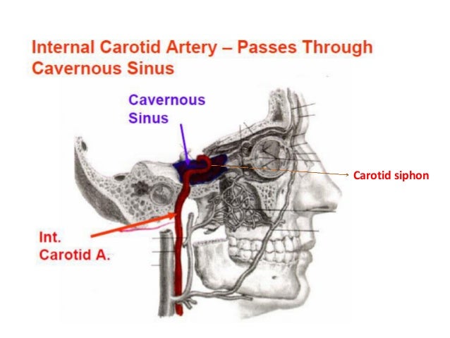
Mar 16, 19 · Most dural arteriovenous fistulas have no clear origin, although some result from identifiable causes such as traumatic head injury, infection, previous brain surgery or tumors Most authorities think that dAVFs involving the larger brain veins usually arise from progressive narrowing or blockage of one of the brain's venous sinuses, whichMar 25, 13 · The cavernous sinuses are a clinically important pair of dural sinuses They are located next to the lateral aspect of the body of the sphenoid bone This sinus receives blood from the superior and inferior ophthalmic veins, the middle superficial cerebral veins, and from another dural venous sinus;Oct 07, 19 · Dural Sinus CVT Anatomy—The dural sinuses are divided into two groups the superior and the inferior dural sinuses The superior dural sinuses comprise the superior sagittal sinus, the inferior sagittal sinus, and the straight sinus (which converge at the confluence of sinuses, or the torcular herophili), and the transverse and sigmoid
Dural Venous Sinuses
Dural venous sinuses labeled
Dural venous sinuses labeled-The occipital sinus is one of the smallest dural venous sinuses and lies, as its name suggests, on the inner surface of the occipital boneTributaries from the marginal sinus of the foramen magnum, some of which connect with both the sigmoid sinus and vertebral venous plexus, coalesce to pass in the attached margin of the falx cerebelli to drain posterosuperiorly at the confluence of theJan 01, 00 · BACKGROUND AND PURPOSE MR venography is often used to examine the intracranial venous system, particularly in the evaluation of dural sinus thrombosis The purpose of this study was to evaluate the use of MR venography in the depiction of the normal intracranial venous anatomy and its variants, to assess its potential pitfalls in the diagnosis of dural venous sinus


15 Dural Venous Sinuses
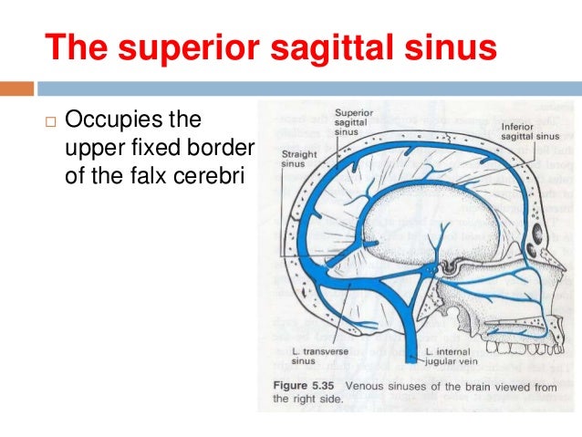
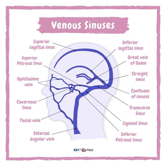
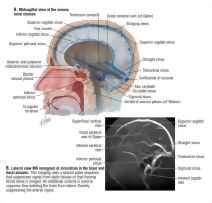
The more downstream venous channels (dural sinus veins) are now visualized — the superior (white) and inferior (dashed white) sagittal sinuses, the Vein of Galen (dashed yellow) and straight sinus (dashed purple), and transverse (dashed red) and sigmoid (dashed blue) sinuses, as well as the inferior petrosal sinus (dashed black)Cranial dural shunts may develop in three distinct areas of the cranial venous system the dural sinuses and their interfaces with bridging veins and emissary veins The exact site of the lesion may dictate the arterial feeders and original venous drainage patternJan 06, 21 · The dural venous sinuses receive cerebral veins from the brain and drain the venous blood mainly into the internal jugular vein (Figures III612 and III613) The superior sagittal sinus is located in the midsagittal plane along the superior aspect of the falx cerebri It drains into the confluence of the sinuses
The dural venous sinuses are clinically important, because you want to AVOID hitting them as they can become a massive source of haemorrhage Yes you can control the bleeding which may eventually cause thrombosis, however the risk of life threatening air embolus is the main problem The large vessel running in the falx cerebri is the superiorAnatomy Dural Sinuses 3 Unpaired Dural Venous Sinuses venous channels found between the endosteal and meningeal layers of dura mater Function is to drain blood in the cranial cavity down to the IJV below the jugular foramen There are paired and unpaired onesAug 01, 16 · Dural Venous Sinuses 1 Superior sagittal sinus lies at the superior attached border of falx cerebri Receives blood from superior cerebral veins (bridging veins) and emissary veins (connects extracranial venous system with intracranial
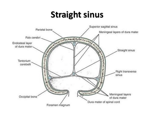
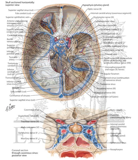
Nov 06, 15 · Dural Venous Sinuses Situated between the layers of the dura mater Dural sinuses are lined by endothelium, and their walls are thick but devoid of muscular tissue They have no valves Function to receive blood from the brain through the cerebral veins and the cerebrospinal fluid from the subarachnoid space through the arachnoid villiJun 17, 21 · The cavernous sinus is the only one of the paired dural sinuses that communicates with each other These sinuses have two intercavernous branches arching over the diaphragma sellae of the pituitary gland;Jan 01, · Extracranial drainage of the posterior cranial fossa dural sinuses, aside from the internal jugular vein, takes place via (1) the anterior condylar vein or the marginal sinus into the internal vertebral venous plexus, (2) the posterior condylar vein into the posterior external vertebral vein, (3) the mastoid emissary, or (4) the occipital


Dural Venous Sinuses Of The Brain Diagram


Dural Venous Sinuses
Dural venous sinus thrombosis (plural thromboses) is a subset of cerebral venous thrombosis, often coexisting with cortical or deep vein thrombosis, and presenting in similar fashions, depending mainly on which sinus is involved As such, please refer to the cerebral venous thrombosis article for a general discussionAnatomy, Imaging and Surgery of the Intracranial Dural Venous Sinuses, 1st Edition Editor R Shane Tubbs This firstofitskind volume focuses on the anatomy, imaging, and surgery of the dural venous sinuses and the particular relevance to neurosurgery and trauma surgery Knowledge of the fine clinical anatomy involved in neurosurgery andThe dural venous sinuses (also called dural sinuses, cerebral sinuses, or cranial sinuses) are venous channels found between the periosteal and meningeal layers of dura mater in the brain


Dural Venous Sinuses


15 Dural Venous Sinuses
By this point, the connection between these sinus wall venous pouches and dural fistulas is hard to miss The prevailing idea is that the site of fistula is in the wall of the sinus, with myriad arteries converging into one or several common venous pouches in sinus walls This is how most simple dural fistulas look likeEvaluation of dural venous sinuses and confluence of sinuses via MRI venography anatomy, anatomic variations, and the classification of variations Childs Nerv Syst 18 Jun;34(6)1111 doi /sCan see small veins coming through the periosteum Brain covered with dura Numerous veins are seen entering the SSS which is faintly seen All dural venous sinuses are situated between two layers of


Main Locations Of Arachnoid Granulations Are Sagittal Sinus Ss Download Scientific Diagram


Dural Venous Sinuses
Jan 01, 17 · All cortical and deep veins drain into the internal jugular veins via special collecting system called dural venous sinuses Dural sinuses are large, endotheliallined trabeculated channels They are placed between periosteal and meningeal layer of dura mater and includes less variations unlike cortical venous system of brainTHEORY OF DURAL VENOUS SINUS VIDEO https//wwwyoutubecom/watch?v=Ebw3OwEQKtcTRANS CRIBRIFORM APPROACH VIDEO https//wwwyoutubecom/watch?v=9v_C94Bwo8s&Dural Venous Sinuses A previously healthy 29yearold female presents with a progressive, diffuse headache and vomiting She has no active illnesses, takes a multivitamin, and an oral contraceptive On exam, there is edema on the scalp, papilledema on fundoscopy, and bilateral muscle weakness Noncontrast head CT shows a hyperdense lesion in a


Dural Venous Sinuses


Dural Venous Sinuses And Veins Of Head And Neck Labeled Eccles Health Sciences Library J Willard Marriott Digital Library
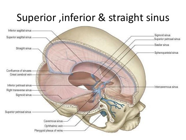
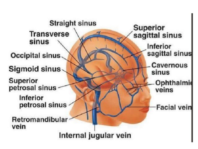
Dural folds made of invaginations of dura mater into crevices of the brain Superior sagittal sinus sigmoid sinus transverse sinus occipital sinus confluence of sinuses inferior sagittal sinus straight sinusDural sinus thrombosis (DST) is a condition where a blood clot forms in one of the dural sinuses The dural sinuses are located in your head They drain deoxygenated blood as well as cerebropsinal fluid (CSF) that surrounds your brain, and empties them into the internal jugular vein that takes it back to your heart and lungs to be reoxygenated They are called venous sinusesDural venous sinuses are venous channels located intracranially between the two layers of the dura mater (endosteal layer and meningeal layer) They can be conceptualised as trapped epidural veins Unlike other veins in the body, they run alone, not parallel to arteries
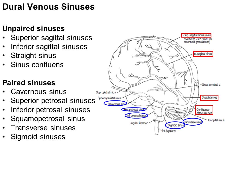


Meninges Brain And Spinal Cord Mbbs Batch 17 Year I Dr Wai Wai Kyi Ppt Download


Cambridge Questions
The dural venous sinuses are spaces between the endosteal and meningeal layers of the dura The sinuses contain an endothelial lining that is continuous into the veins that are connected to themJul 31, 19 · Dural Venous Sinuses Of The Brain Diagram Dural Venous Sinuses Of The Brain Diagram In this image, you will find superior sagittal sinus, falx cerebri, inferior sagittal sinus, straight sinus, cavernous sinus, transverse sinuses, sigmoid sinus, jugular foramen, right internal jugular vein in it Our SECOND youtube film is ready to runID Title Dural Venous Sinuses Category LabeledAnatomy Atlas 6E ID Category LabeledCochard Imaging 1E Radiographs ID Title Dural Venous Sinuses Category LabeledHansen FC 3E
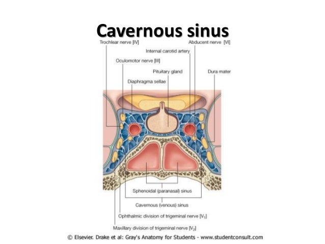


Ehcahyp Od6aym


The Cranial Cavity Contents 1 The Brain And
Feb 16, 17 · Dural venous sinuses are venous channels that are present usually the two layers of dura mater They are lined by endothelium They do not have muscle inAnatomy Image Database Dural Venus Sinuses Skull cap The location of the superior sagittal sinus is faintly visible Why is the sinus not seen?Mar 01, 16 · Characteristic feature of dural venous sinuses • Lined by endothelium, no muscular coat & valveless • Collect blood from brain,meninges, orbit,internal ear & diploe • Connected to valveless emissary veins to maintain the internal & external venous pressure • Projection of arachnoid granulation into it for CSF absorption


Pin On Nbde Part I
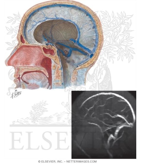


Dural Venous Sinuses
BACKGROUND AND PURPOSE The suboccipital cavernous sinus, a vertebral venous plexus surrounding the horizontal portion of the vertebral artery at the skull base, provides an alternative pathway of cranial venous drainage by virtue of its connections to the cranial dural sinuses, the vertebral venous plexus, and the jugular venous systemJun 16, 21 · The anatomical diversity in the dural venous sinuses is significant The practicing neuroradiologist must have a thorough understanding of dural venous sinus architecture and typical anatomic variations to distinguish between normal and pathologic situations We use an imagebased method to evaluate dural venous sinus architecture as well as highlight frequentDural venous sinuses venous channels found btwn endosteal and meningeal layers of dura mater fx drain venous blood w/in cranial cavity to CV via internal jugular vein paired or unpaired w/ most near falx cerebri or cavernosus sinus (only great cerebral v and cavernous sinus aren't)
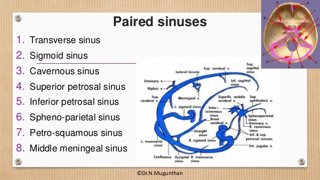


Dural Venous Sinuses Cavernous Sinus Dr N Mugunthan


Dural Venous Sinuses Learning Anatomy Facebook
The torcular Herophili (confluence of sinuses) is a highly variable structure that constitutes a vital part of the network of dural venous sinuses The torcular Herophili is defined historically as the intersection of the superior sagittal sinus, the straight sinus, the occipital sinus, and the two transverse sinusesDural venous sinuses There are seven paired (transverse, cavernous, greater & lesser petrosal, sphenoparietal, sigmoid and basilar) and five unpaired (superior & inferior sagittal, straight, occipital and intercavernous) dural sinusesMay 31, 21 · Confluence of Sinuses (confluens sinuum) The confluence of sinuses (torcular of Herophilus or torcula) is a dural venous sinus that appears as a dilation at the posterior end of the superior sagittal sinusIt is located on the caudal aspect of the brain, around the internal occipital protuberance of the occipital bone Functionally, this vessel represents a junction between the


Dural Venous Sinuses 3d Anatomy Tutorial Youtube


Dural Venous Sinuses Diagram Quizlet
The dural venous sinuses can be either paired or unpaired Paired sinuses include the transverse sinus, cavernous sinus, superior petrosal sinus, inferior petrosal sinus, sphenoparietal sinus, andAnatomy and Function of the Dural Venous Sinuses See online here The dural venous sinuses (DVS) are venous blood reservoirs located between the 2 layers of the dura mater The absence of lymphatic drainage in the brain places the venous outflow system means that the DVS isMar 16, 19 · Treatment Neurosurgery for dural arteriovenous fistulas Mayo Clinic neurosurgeons review surgical options for a dural arteriovenous fistula Endovascular procedures In an endovascular procedure, your doctor may insert a long, thin tube (catheter) into a blood vessel in your leg or groin and thread it through blood vessels to the dural


Dural Venous Sinuses


Anatomy Of The Cavernous Sinus Anatomy Drawing Diagram
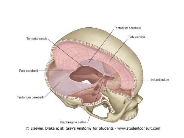
Cerebral venous sinus thrombosis (CVST), cerebral venous and sinus thrombosis or cerebral venous thrombosis (CVT), is the presence of a blood clot in the dural venous sinuses (which drain blood from the brain), the cerebral veins, or bothSymptoms may include severe headache, visual symptoms, any of the symptoms of stroke such as weakness of the face and limbs on one side ofOne anterior and the other posterior to the infundibulum They drain posteriorly by the petrosal sinusesDural venous channels — venous sinuses in the tentorium cerebelli that connect cerebellar and inferior cortical veins to the named venous sinuses (transverse, sigmoid) are well known There is surgical literature on being careful not to cut them and risk venous infarction
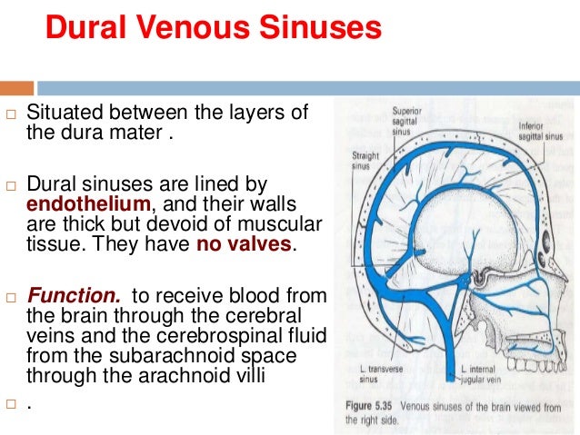


15 Dural Venous Sinuses
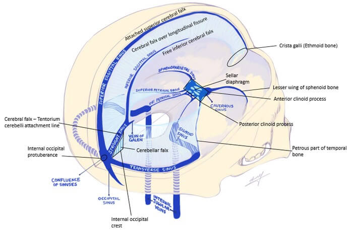


Dural Reflections And Venous Sinuses Epomedicine
This video provides a walkthrough of the dural venous sinuses (eg transverse sinus, cavernous sinus) You can follow along using our free written guide witIn this tutorial we will review the anatomy and configuration of the dural venous sinuses We will be learning about the folllowing structures Superior sagMar 09, 16 · The dural venous sinuses lie between the periosteal and meningeal layers of the dura mater They are best thought of as collecting pools of blood, which drain the central nervous system, the face, and the scalp All the dural venous sinuses ultimately drain


Grooves For Dural Venous Sinuses Paranasal Sinuses Diagram Quizlet


Cardiovascular Lab Veins Of The Dural Venous Sinuses Diagram Quizlet
The dural entrance of cerebral bridging veins into the superior sagittal sinus an anatomical comparison between cadavers and digital subtraction angiography Han H(1), Tao W, Zhang M Author information (1)Department of Anatomy, Anhui Medical University, Hefei, China


Straight Sinus Ultrastructural Analysis Aimed At Surgical Tumor Resection In Journal Of Neurosurgery Volume 125 Issue 2 16
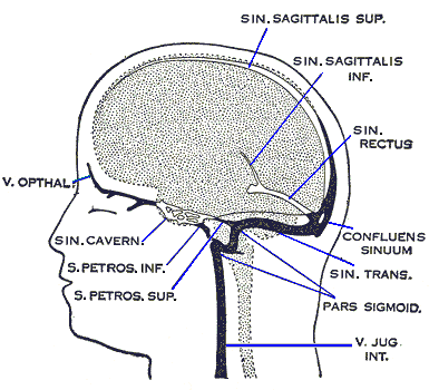


Anatomy And Function Of The Dural Venous Sinuses Medical Library


Dural Venous Sinuses Summaries For Medical Students


Cerebral And Sinus Vein Thrombosis Circulation


Dural Venous Sinuses Radiology Reference Article Radiopaedia Org
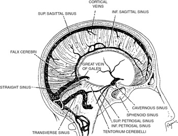


Venous And Dural Sinus Thrombosis Chapter 13 Vertebrobasilar Ischemia And Hemorrhage
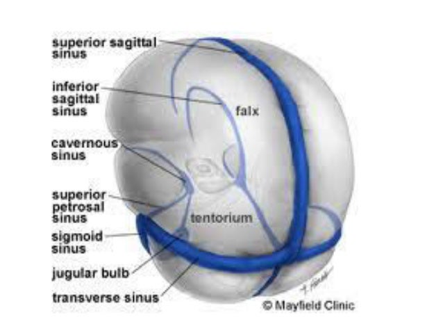


Ehcahyp Od6aym


Dural Venous Sinuses An Overview Sciencedirect Topics
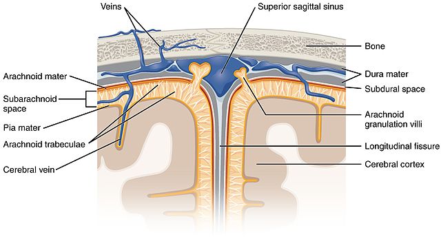


Anatomy And Function Of The Dural Venous Sinuses Medical Library
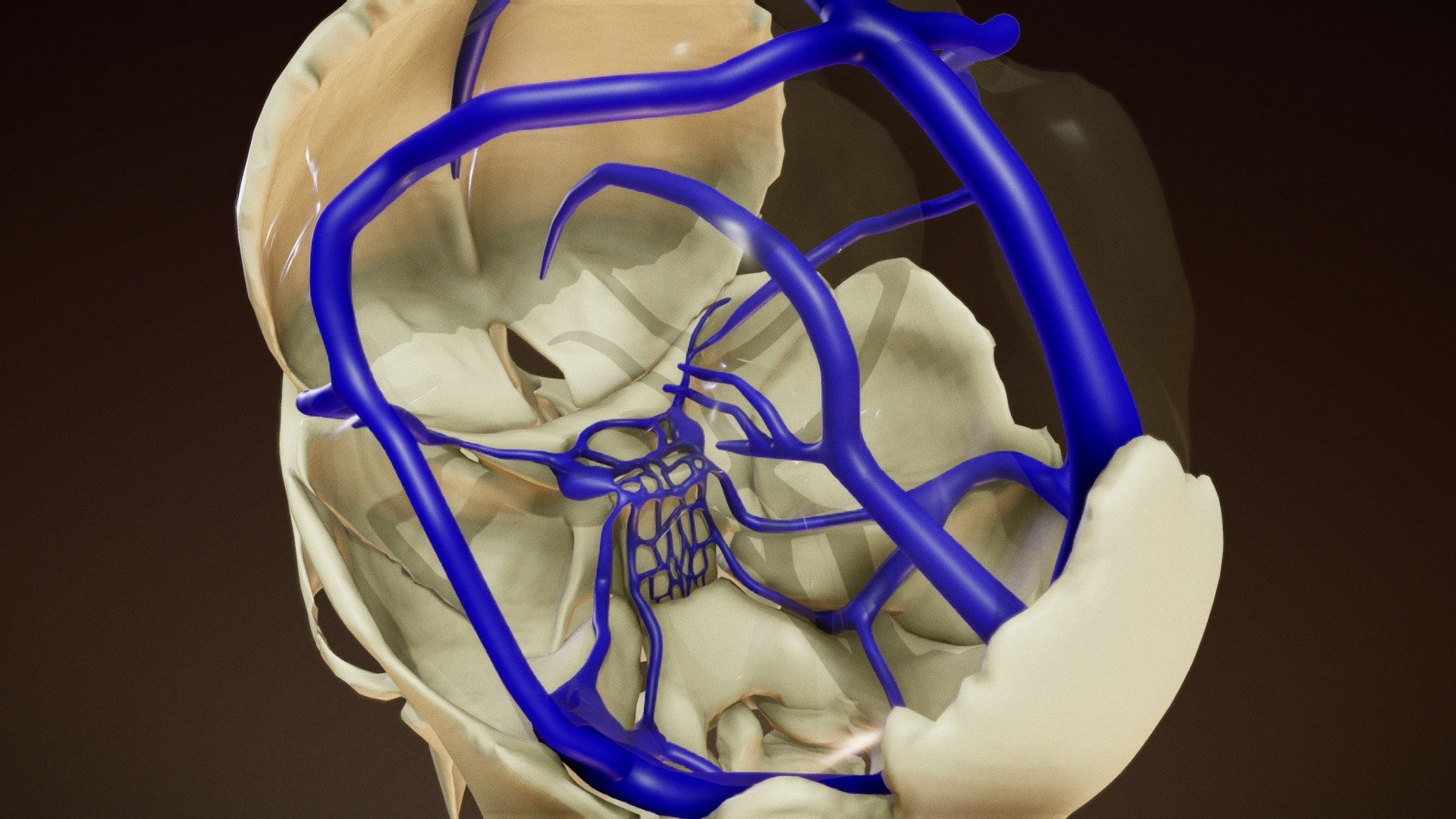


Dural Venous Sinuses Buy Royalty Free 3d Model By Anatomy By Doctor Jana Docjana 07c7b


Pdf Anatomy Of Cerebral Veins And Sinuses


Dural Venous Sinuses


1


Venous Sinuses Neuroangio Org
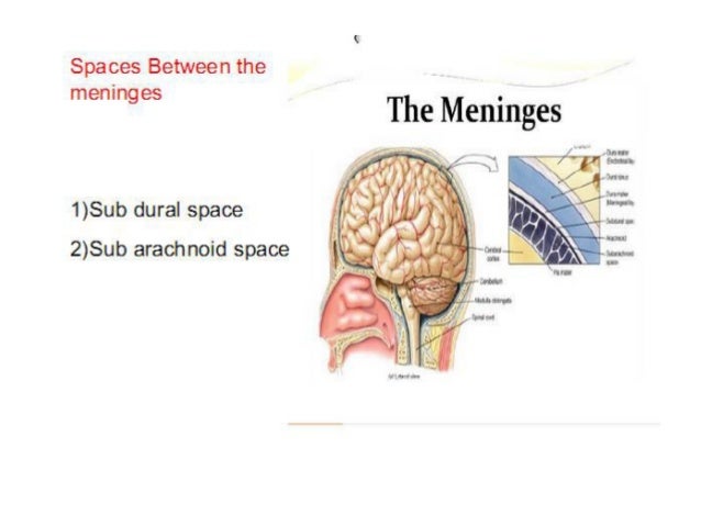


Dural Venous Sinuses


Dural Venous Sinuses Diagram Quizlet


Dural Venous Sinuses


Dural Venous Sinuses Anatomy Kenhub


31 Ot Neuroscience Ideas Anatomy And Physiology Brain Anatomy Neuroscience


Pin On Brain


Dural Venous Sinuses An Overview Sciencedirect Topics
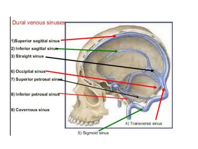


Dural Venous Sinuses


Dural Venous Sinuses
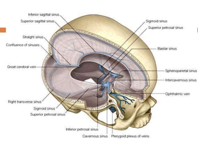


15 Dural Venous Sinuses


Dural Venous Sinuses Youtube


Dural Venous Sinuses Diagram Quizlet
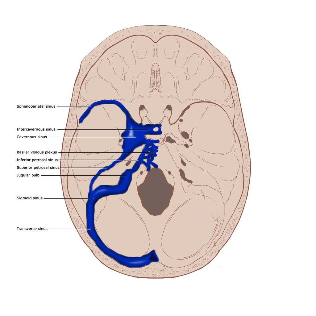


Dural Venous Sinuses Illustration Radiology Case Radiopaedia Org


Dural Venous Sinuses 3d Anatomy Tutorial Youtube


Trying To Visualize The Dural Venous Sinuses Diagnostic Medical Sonography Arteries Anatomy Brain Anatomy


The Dural Venous Sinuses Rapid Review Youtube


Cerebral And Sinus Vein Thrombosis Circulation
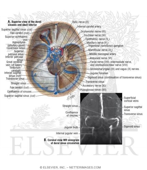


Dural Venous Sinuses


Superior Sagittal Sinus Wikipedia
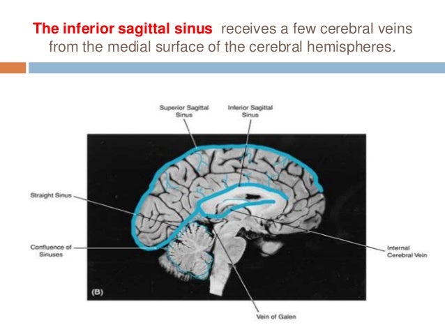


15 Dural Venous Sinuses


Dural Venous Sinuses Diagram Quizlet
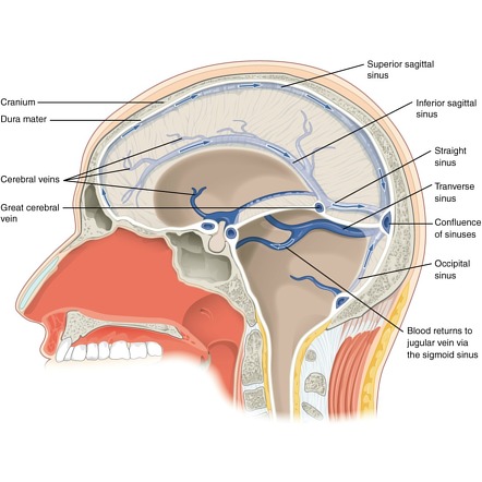


Dural Venous Sinuses Radiology Reference Article Radiopaedia Org


Dural Venous Sinuses Radiology Reference Article Radiopaedia Org
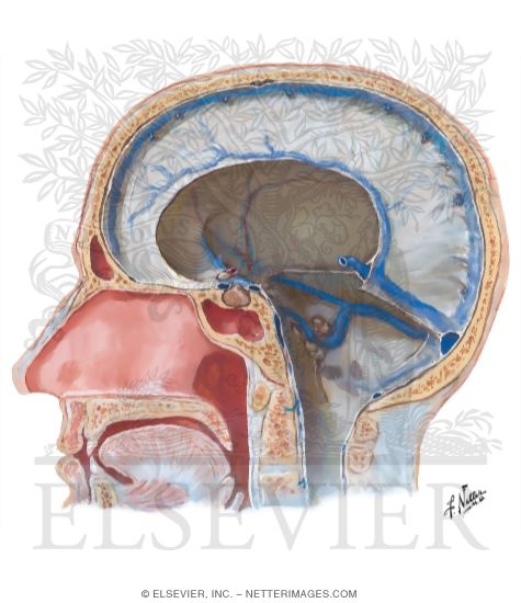


Dural Venous Sinuses


Http Www Headandnecktrauma Org Wp Content Uploads 16 10 Meninges And Dural Sinuses Pdf


1


Dural Venous Sinuses An Overview Sciencedirect Topics


Pin On Psychology
:background_color(FFFFFF):format(jpeg)/images/library/13675/meninges-and-arachnoid-granulations_english.jpg)


Superior Sagittal Sinus Anatomy Tributaries Drainage Kenhub


1


Dural Venous Sinuses


Dural Venous Sinuses Ventricles Diagram Quizlet
:background_color(FFFFFF):format(jpeg)/images/library/13677/8iVwiSwwNxvJhpSQELoQ_Sinus_sagittalis_superior_02.png)


Superior Sagittal Sinus Anatomy Tributaries Drainage Kenhub


Dural Venous Sinuses Diagram Quizlet
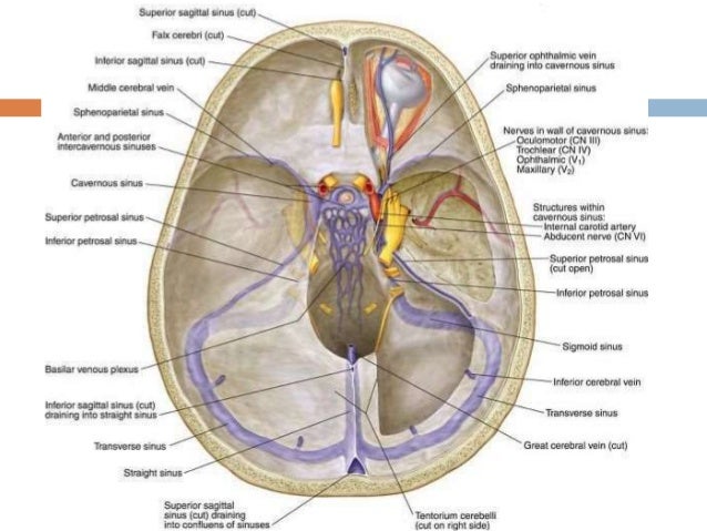


15 Dural Venous Sinuses


Dural Venous Channels Neuroangio Org
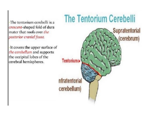


Dural Venous Sinuses


Dural Venous Sinuses
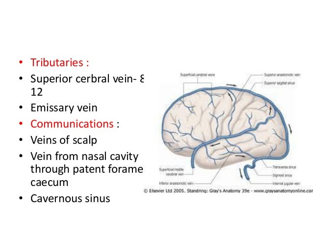


Dural Venous Sinuses


Mrv Major Venous Sinuses Are Labeled With Arrows Download Scientific Diagram


Dural Venous Sinuses Anatomy Anatomy Drawing Diagram
:background_color(FFFFFF):format(jpeg)/images/article/en/dural-sinuses/MBT9Z5mR2o8TnY2hb131w_Superficial_veins_of_the_brain_lateral_view.png)


Dural Venous Sinuses Anatomy Kenhub


Dural Venous Sinuses Wikipedia


Venous Sinuses Of The Brain Google Search Con Imagenes Anatomia Medica
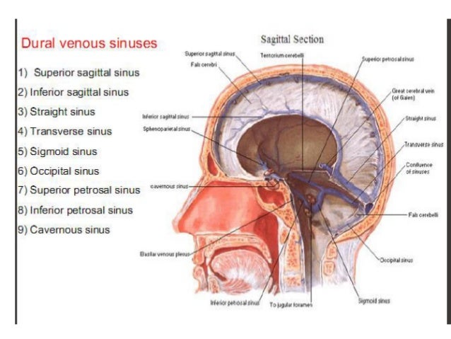


Dural Venous Sinuses


File Dural Sinuses Jpg Wikimedia Commons


Dural Venous Sinuses Wikipedia
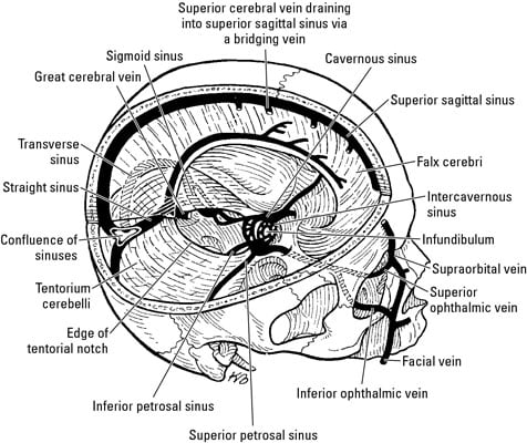


Anatomy Of The Brain The Meninges Dummies
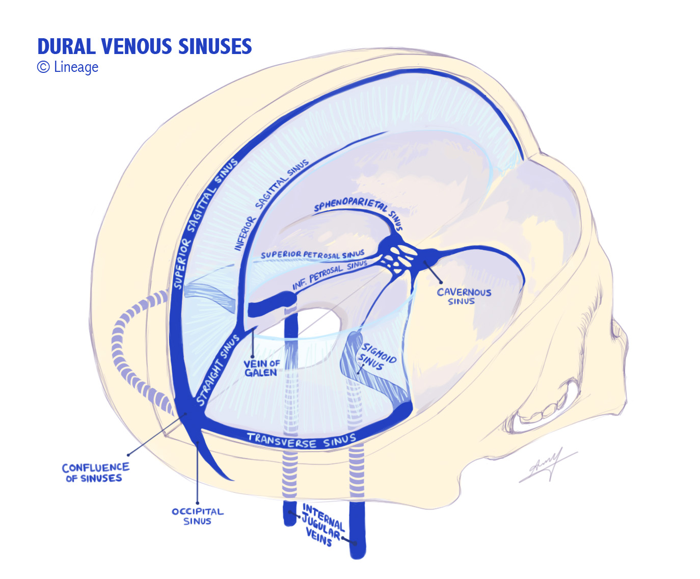


Dural Venous Sinuses Medmule
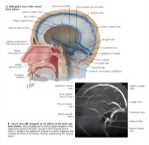


Dural Venous Sinuses
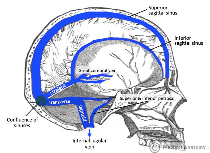


Venous Drainage Of The Cns Cerebrum Teachmeanatomy
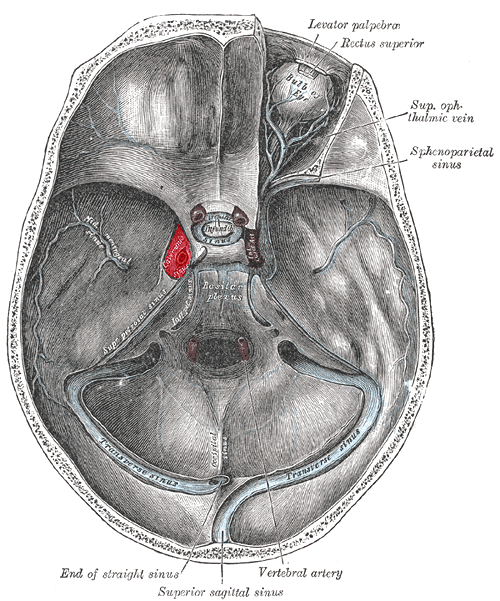


Cavernous Sinus Wikiwand
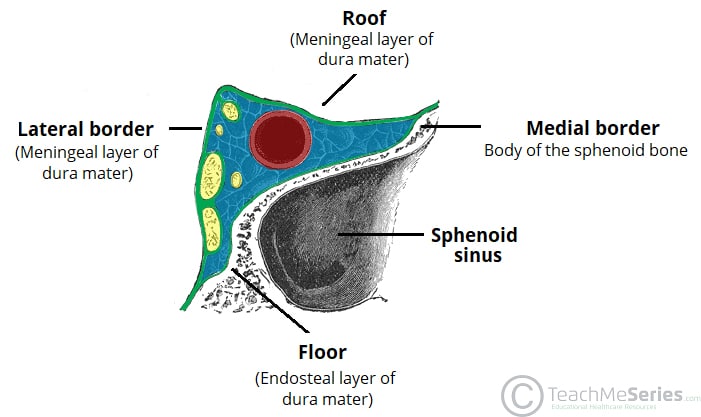


The Cavernous Sinus Contents Borders Thrombosis Teachmeanatomy


Dural Venous Sinuses


Dural Venous Sinuses


Venous Sinuses Neuroangio Org


Ehcahyp Od6aym


Dural Venous Sinuses


1


Venous Sinuses Neuroangio Org


Dural Venous Sinuses Diagram Quizlet
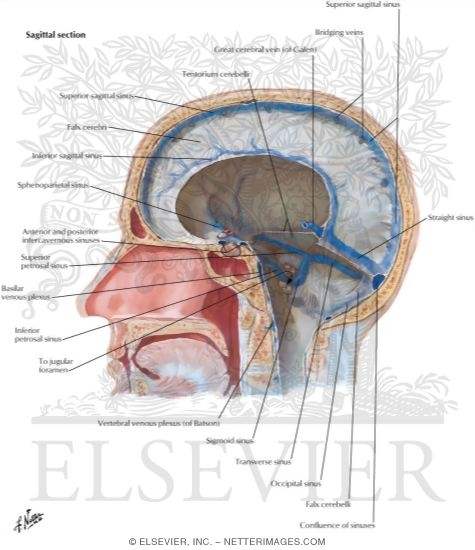


Dural Venous Sinuses
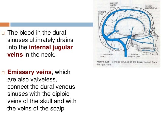


15 Dural Venous Sinuses
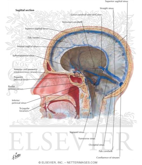


Dural Venous Sinuses
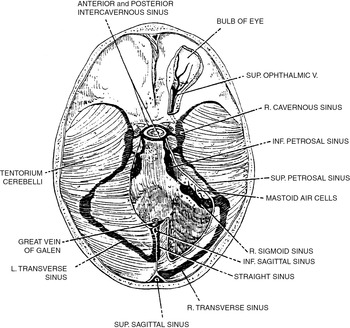


Venous And Dural Sinus Thrombosis Chapter 13 Vertebrobasilar Ischemia And Hemorrhage


Dural Venous Sinuses


コメント
コメントを投稿